

Abstract
Background This study retrospectively evaluated the clinical outcomes of intramedullary nailing of femoral shaft fractures with third fragments and analyzed the risk factors for delayed union.
Methods Retrospective analyses involving 51 patients who underwent intramedullary nailing of femoral shaft fractures with third fragments (AO classification type B, 35 cases; type C, 16 cases) were conducted. Delayed union was defined as either more than 10 months required for callus formation in more than three of the four cortical bone surfaces observed in the frontal and lateral radiographic views or the requirement for additional surgery such as nail conversion or bone transplantation. Seventeen patients developed delayed union (D group). Thirty-four patients achieved bony union within 9 months (U group). The following background variables were compared between groups: age at the time of the injury; AO classification; ratio of open fracture; waiting period before surgery; rate of the infraisthmal fracture; diameter of the intramedullary nail; ratio of the intramedullary nail to the femur; length and displacement of the third fragment; and use of open reduction, poller screws, or dynamization.
Results Significant differences were found between the D and U groups for age (32.2±14.1 vs. 25.3±9.6 years), open fracture ratio (35.3% vs. 11.8%), and displacement of the third fragment (13.7±6.4 vs. 9±6.3 mm). Multiple logistic regression analysis only identified displacement of the third fragment as a risk factor for delayed union (p=0.03; OR 1.13; 95% CI 1.01 to 1.26).
Discussion Delayed union was observed in 17 cases (33.3%) after intramedullary nailing of femoral shaft fractures with third fragments. Displacement of the third fragment influenced delayed union.
Level of evidence Level III.
Introduction
Intramedullary nailing (IMN) is currently considered the gold standard for femoral shaft fractures (AO classification 32). Much has been reported regarding its favorable bony union rate and lower complication rates.1–7 However, delayed union or non-union after IMN occasionally occurs.8–12 These complications severely affect the activities of daily living and cause socioeconomic problems. Several risk factors for delayed union have been detected, such as AO classification type C, infraisthmal fracture, and not using the reaming technique. However, a few reports have focused on the third fragment. Furthermore, the perception of whether it is better to use open reduction for the displacement of the third fragment is limited. If risk factors become evident, then developing a treatment strategy for comminuted fractures is helpful for reducing the suffering caused by these complications. This study retrospectively evaluated the clinical outcomes of IMN for femoral shaft fractures with third fragments and analyzed risk factors for delayed union.
Patients and methods
Patient characteristics
Between 2008 and 2015, a total of 105 skeletally mature patients with IMN of femoral shaft fractures underwent treatment. Eighty-nine patients were included as study candidates after excluding those with subtrochanteric fractures (less than 5 cm distal from the lesser trochanter) and double fractures (third fragment presented with a cylindrical shape). Among them, 74 patients were able to be followed up for more than 1 year (follow-up rate, 83.1%). We recruited 51 patients who underwent IMN of femoral shaft fractures with third fragments (AO classification type B, 35 cases; type C, 16 cases), including one patient with an ipsilateral femoral neck, one with a femoral condyle, and one with a lower leg fracture. Two additional patients had contralateral proximal femoral fractures. There were 44 male patients (86.3%) and 7 female patients (13.7%). Mean age at the time of the injury was 27.6±11.6 years (range, 14–61). There were 10 (19.6%) open fractures (Gustilo-Anderson classification type 1, four cases; type 2, six cases), but soft tissue reconstruction was not necessary for all cases. Motor vehicle crash was the most common cause of trauma (43 patients). Five patients were injured by falling from a height. One patient was injured in a skiing accident. One patient was injured in a hit-and-run motor vehicle crash with a truck. One patient was run over by a train. The mean follow-up period was 22±10 months (range, 12–58). Exclusion criteria were pathological fractures and a history of femur surgery.
Surgical techniques
Surgery was performed with the affected limb in traction on a fracture reduction table with the patient in the supine position. Antegrade IMN was performed for all cases (T2 Recon Nailing System; Stryker, Kalamazoo, MI, USA). Reaming was performed for 49 cases; two cases did not undergo reaming. Proximal locking screws were used in the antegrade femoral mode for 40 cases, and the recon mode was used for 11 cases. Distal locking screws with both static and dynamic holes were used. Poller screws13 were used for 10 cases. Open reduction of the third fragment was performed simultaneously with IMN for 13 cases. Of these, cerclage wiring was performed for five cases. Additional surgery for fracture union was necessary for eight cases; nail conversion for four cases, a free bone graft for three cases, and a vascularized fibula graft for one case.
Definitions of delayed union
Delayed union was defined as either more than 10 months for callus formation in more than three of the four cortical bone surfaces observed in the frontal and lateral radiographic views or the requirement for additional surgery.
Evaluations
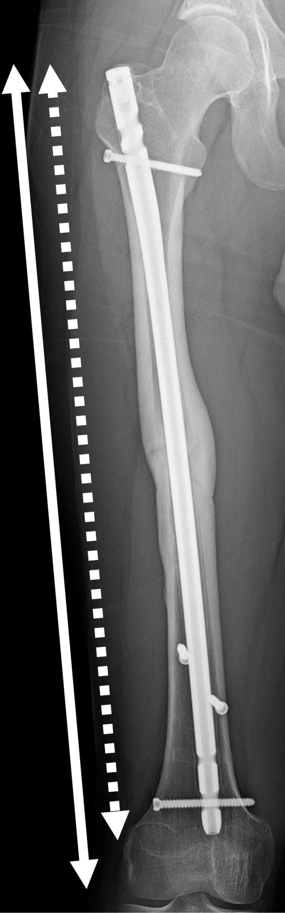
First, patients were classified into two groups. Seventeen patients who developed delayed union constituted the D group. Thirty-four patients who achieved bony union within 9 months constituted the U group. The following background variables were compared between groups: age at the time of injury; AO classification; open fracture ratio; waiting period before surgery; infraisthmal fracture rate; nail diameter; nail-to-femur ratio (figure 1); length of the third fragment (figure 2; the average value of the measured longitudinal length of the third fragment in the frontal and lateral radiographic views); displacement of the third fragment (figure 3; the maximum value of the measured space between the cortical bone surface of the shaft and third fragment in the frontal and lateral radiographic views); and the use of open reduction, poller screws for infraisthmal fracture, or dynamization (removal of the distal locking screws as planned and accidental breakage of the locking screws). Second, the following background variables were compared for 13 patients who underwent open reduction: AO classification; length of the third fragment; and displacement of the third fragment.

Nail-to-femur ratio. This was calculated as the ratio of the nail length (dotted line) to the femoral length (solid line).
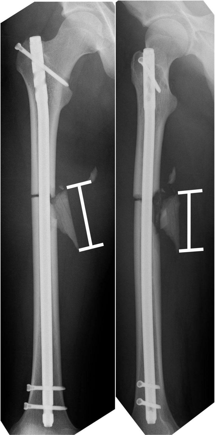
Length of the third fragment. This was the average value of the measured longitudinal lengths of the third fragment in the frontal (left) and lateral (right) radiographic views.
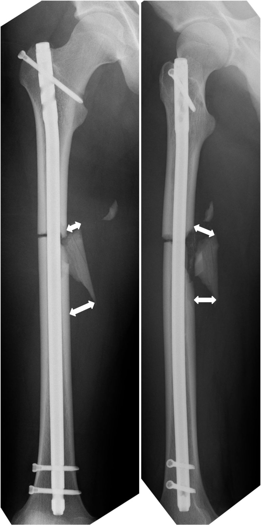
Displacement of the third fragment. This was the maximum value of the measured space (total of 4 points) between the cortical bone surface of the shaft and the third fragment in the frontal (left) and lateral (right) radiographic views.
Statistical analyses
Statistical analyses were performed using SPSS Statistics software (V.22; IBM). Variables were compared between the two groups using the Student’s t-test or χ2 test, and p<0.05 was considered significant. Furthermore, we analyzed the risk factors for delayed union or non-union using a multiple logistic regression analysis.
Results
There were significant differences between age at the time of injury (32.3±14.1 vs. 25.3±9.6 years; p=0.04), ratio of open fractures (6 cases [35.3%] vs. 4 cases [11.8%]; p=0.05), and displacement of the third fragment (13.7±6.4 vs. 9±6.3 mm; p=0.02) for all patients in the D group and U group. There were no significant differences between groups regarding the AO classification (type B: 10 vs. 25 cases; type C: 7 vs. 9 cases; p=0.29), waiting period before surgery (3.8±7.8 vs. 2.1±4 days; p=0.30), rate of infraisthmal fractures (9 cases [52.9%] vs. 17 cases [50%]; p=0.84), nail diameter (10.8±1.4 vs. 10.1±1.3 mm; p=0.10), nail-to-femur ratio (90.4%±0.05% vs. 90.3±0.03%; p=0.95), length of the third fragment (90.3±46 vs. 77±40.8 mm; p=0.30), open reduction (6 cases [35.3%] vs. 7 cases [20.6%]; p=0.26), poller screws (4 cases [23.5%] vs. 6 cases [17.6%]; p=0.62), and dynamization (7 cases [41.2%] vs. 13 cases [38.2%]; p=0.84) (table 1).
Clinical results of all patients in the D group and U group
For 13 patients who underwent open reduction, there were no significant differences between delayed union and normal union for displacement of the third fragment (9.7±4.3 vs. 10.3±6.1 mm; p=0.85). However, the ratio of AO classification type C (4 cases [66.7%] vs. 1 case [14.3%]; p=0.05) and the length of the third fragment (105.1±24 vs. 72.7±21 mm; p=0.02) were significantly different (table 2).
Clinical results of 13 patients who underwent open reduction
The multiple logistic regression analysis showed that only displacement of the third fragment significantly affected delayed union (p=0.03; OR 1.13; 95% CI 1.01 to 1.26); however, age (p=0.13), AO classification (p=0.19), and open fracture (p=0.17) did not (table 3).
Multiple logistic regression analysis of risk factors for delayed union
Discussion
We conducted retrospective analyses of 51 patients who underwent IMN of femoral shaft fractures with third fragments. As a result, there were significant differences between the D group and U group for age at the time of injury, ratio of open fractures, and displacement of the third fragment. The main finding of this study was that the multiple logistic regression analysis showed that only displacement of the third fragment significantly affected delayed union.
The non-union rate after IMN of femoral shaft fractures ranges from 1% to 20%, depending on the type of fracture and surgical technique.14 At our institution, we treated 23 cases of AO classification type A at approximately the same time, and only one case developed into delayed union. However, the remaining 22 cases (95.7%) showed complete healing. Therefore, we limited our research to AO classification types B and C and excluded type A. As a result, delayed union was observed in 17 cases (33.3%). It was suggested that delayed union was not a negligible complication in cases of femoral shaft fractures with third fragments.
Several risk factors for delayed union or non-union have been detected, such as AO classification type C,15 16 length, displacement of the third fragment,17 18 and not using the reaming technique.19–21 Watanabe et al22 reported open fractures, infraisthmal fractures, breakage of the locking screw, and inappropriate dynamization as risk factors for non-union after IMN. They recommended a combination of poller screws and, if possible, a larger diameter nail to improve the stability of the infraisthmal femoral fracture. In general, intervention could not be performed for factors related to fracture characteristics themselves, such as AO classification, open fractures, infraisthmal fractures, and length of the third fragment. However, intervention could be performed for factors related to surgical techniques. In this study, the nail diameter was more than 10 mm, and the nail-to-femur ratio was more than 90% for both groups. Therefore, nails of an appropriate size were selected. Furthermore, reaming was performed for 49 (96%) of 51 patients, with or without poller screws, and dynamization did not differ in both groups. Therefore, factors related to surgical techniques were considered to be eliminated.
Regarding the fracture characteristics, only displacement of the third fragment could undergo intervention with intraoperative reduction. In this study, displacement of the third fragment was significantly larger in the D group, and multiple logistic regression analysis indicated that displacement of the third fragment was the only significant factor. Lin et al17 reported that the union rates of the small-gap (≤10 mm) and large-gap (>10 mm) groups were 75.9% and 21.1%, respectively. They concluded that larger displacement of the fragment could indicate a worse environment resulting from potential soft tissue interposition and poor axial load-bearing ability. Lee et al18 reported that non-union developed significantly more frequently with fragments 8 cm or longer or when the displacement was 20 mm or more in the proximal area and 10 mm or more in the distal area. We agreed with their perception that the degree of displacement has more influence on the union rate than the third fragment size. It was suggested that reduction of the third fragment is important. According to our results, displacement ≤10 mm is one possible reference standard for avoiding delayed union.
Approximately 10 mm of displacement remaining despite open reduction was considered to be due to cerclage wiring only being performed for only five cases in this study. However, whether open reduction and cerclage wiring are effective for bony union is a controversial topic. In this study, 6 of 13 cases that underwent open reduction developed into delayed union. In contrast to seven cases that achieved bony union, there were no significant differences in displacement of the third fragment; however, the ratio of AO classification type C and the length of the third fragment were significantly different. Damage to the surrounding soft tissues, such as the periostea, muscles, and vessels, was suspected to be worse in these fracture patterns, and additional damage due to open reduction itself could have adversely affected bony union. Burç et al23 reported that for 44 patients with femoral shaft fractures with open reduction, the complete union rate was 90.9% for 40 patients; the non-union rate was 9.1% for four patients who underwent IMN. They concluded that the open technique was acceptable because the results of their study were similar to the results of the closed IMN technique in the literature. However, their study was composed of 34 patients with AO classification type A and 10 patients with type B. It was not mentioned whether non-union cases were observed for type A or type B, but the results were considered difficult to accept. Therefore, further investigations must be performed to determine whether open reduction is effective for bony union.
Wagner24 mentioned the locking compression plate (LCP) as an alternative to IMN and other fixation techniques, especially for cases of multifragmentary shaft and metaphyseal fractures of the femur. Apivatthakakul and Chiewcharntanakit25 reported good clinical results for 26 patients who achieved complete bony union; only 2 of 28 patients treated with the minimally invasive plate osteosynthesis (MIPO) technique using a broad dynamic compression plate had delayed union (AO classification type B, 13 patients; type C, 15 patients). Wenda et al26 similarly recommended the MIPO technique for comminuted femoral shaft fractures. We used IMN as our first choice of treatment for femoral shaft fractures. However, application of the LCP with the MIPO technique might be considered for AO classification types B and C, depending on the length and displacement of the third fragments.
This study had several limitations. First, only a small number of patients were included. Second, this was not a prospective study. Larger scale prospective studies should be conducted so that rigorous analyses can be performed. Third, different surgeons had performed the operations. However, we think the risk factors related to fracture characteristics themselves could be rigorously evaluated with minimum bias because unified nails were used for all cases, and there were no significant differences between groups regarding the waiting period, nail diameters, nail-to-femur ratio, or the use of open reduction, poller screws, or dynamization, which were all related to surgical techniques.
In summary, we used IMN as our first choice of treatment, even for comminuted femoral shaft fractures. However, delayed union rates were not low, especially for fractures with larger and more displaced third fragments. Surgeons should try to reduce displacement so that improved clinical results can be achieved for these fracture patterns.
Conclusions
We retrospectively evaluated the clinical outcomes of IMN of femoral shaft fractures with third fragments and analyzed the risk factors for delayed union observed in 17 cases (33.3%). Significant differences were found for age at the time of injury, ratio of open fractures, and displacement of the third fragment. The main finding of this study was that, according to the multiple logistic regression analysis, only displacement of the third fragment significantly affected delayed union. According to our results, displacement ≤10 mm is one possible reference standard for avoiding delayed union. If surgeons predict that displacement of the third fragment ≤10 mm cannot be achieved using a closed maneuver, then open reduction using a bone clamp or cerclage wire should be considered.
Acknowledgments
We thank the reviewers for their valuable comments and suggestions. We also thank our trauma team and the junior and senior residents for their helpful contributions to our study.
Footnotes
Contributors KH, GE, and TU performed the surgery. KH, YU, and YK collected the data and drafted the article. TU led the statistical analyses. YU and MW revised the article. All authors read and approved the final article.
Funding The authors have not declared a specific grant for this research from any funding agency in the public, commercial or not-for-profit sectors.
Competing interests None declared
Patient consent for publication Obtained.
Ethics approval This study was approved by the Institutional Review Board for Clinical Research of Tokai University School of Medicine (No 17R215). All patients received information regarding the purpose prior to being included in this study.
Provenance and peer review Not commissioned; externally peer reviewed.
This is an open access article distributed in accordance with the Creative Commons Attribution Non Commercial (CC BY-NC 4.0) license, which permits others to distribute, remix, adapt, build upon this work non-commercially, and license their derivative works on different terms, provided the original work is properly cited, appropriate credit is given, any changes made indicated, and the use is non-commercial. See: http://creativecommons.org/licenses/by-nc/4.0/.


